Equine Diagnostic Imaging
Specialised X-Ray, Ultrasound and Edoscopic Services for Horses
Equine Diagnostic Imaging
Specialised X-Ray, Ultrasound and Edoscopic Services for Horses
Diagnostic Imaging
Radiography and ultrasound are the most commonly used imaging techniques in horses and our vets have access to a full range of portable digital equipment.
All images obtained from these techniques are stored digitally and are available immediately to our veterinarians.

Radiography (X-Ray)
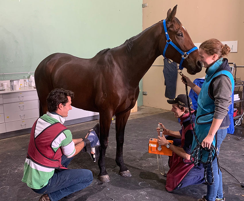
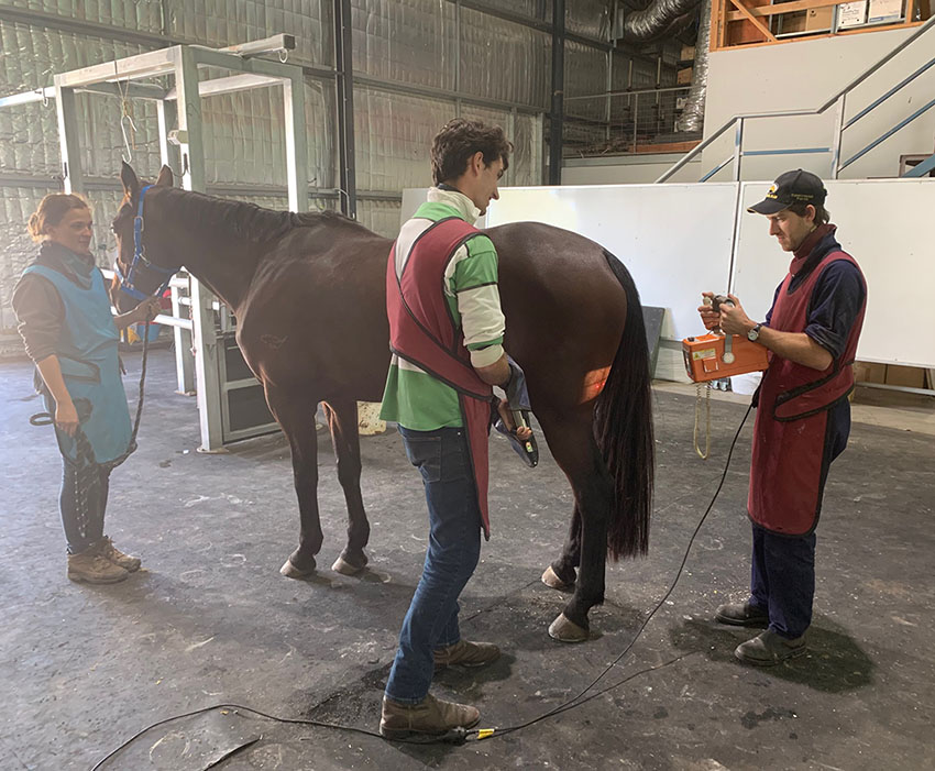
Radiography is the most commonly used imaging technique used to evaluate lameness by allowing us to visualise bones and joints. Our portable digital radiography unit allows us to assess the lower limb, upper limbs, head, neck and the back. Some areas of the horse eg pelvis and lower back are not able to be adequately imaged with radiography. In these cases other imaging modalities may be used including ultrasound.

Ultrasonography (Ultrasound)
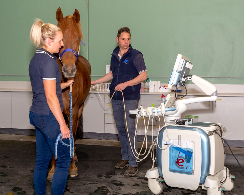
Ultrasound is used for the diagnosis of musculoskeletal conditions including joint, tendon, muscle and ligament injuries and it is effective for monitoring the healing process in these cases. In addition to this we also use ultrasound for evaluation of horses with abdominal pain (colic), respiratory disease and for cardiology examinations. Ultrasound is also routinely used to monitor broodmare reproductive cycles, for equine pregnancy detection (including twinning), and late pregnancy.
We have a high quality hospital based machine which is excellent for scanning more difficult areas such as the chest, heart and abdomen. We also have a portable ultrasound machine which is predominantly used for reproductive examinations.

Equine Endoscopy
Videoendoscopy allows visualisation of the horse’s upper respiratory tract, urinary tract and parts of the gastrointestinal tract. The endoscopes are linked to a screen which allows the vets and the clients to see the images at the same time and for the images to be recorded.
We have a range of different sized endoscopes that we can use to visualise these different regions of the horse in great detail and take biopsy samples for laboratory diagnosis.
Endoscopy of the stomach (gastroscopy) and duodenum (gastroduodenoscopy) is commonly performed in horses that have a history of a poor appetite, recurrent bouts of abdominal pain, behavioral complaints and weight loss.
Standing MRI at South Eastern Equine Hospital
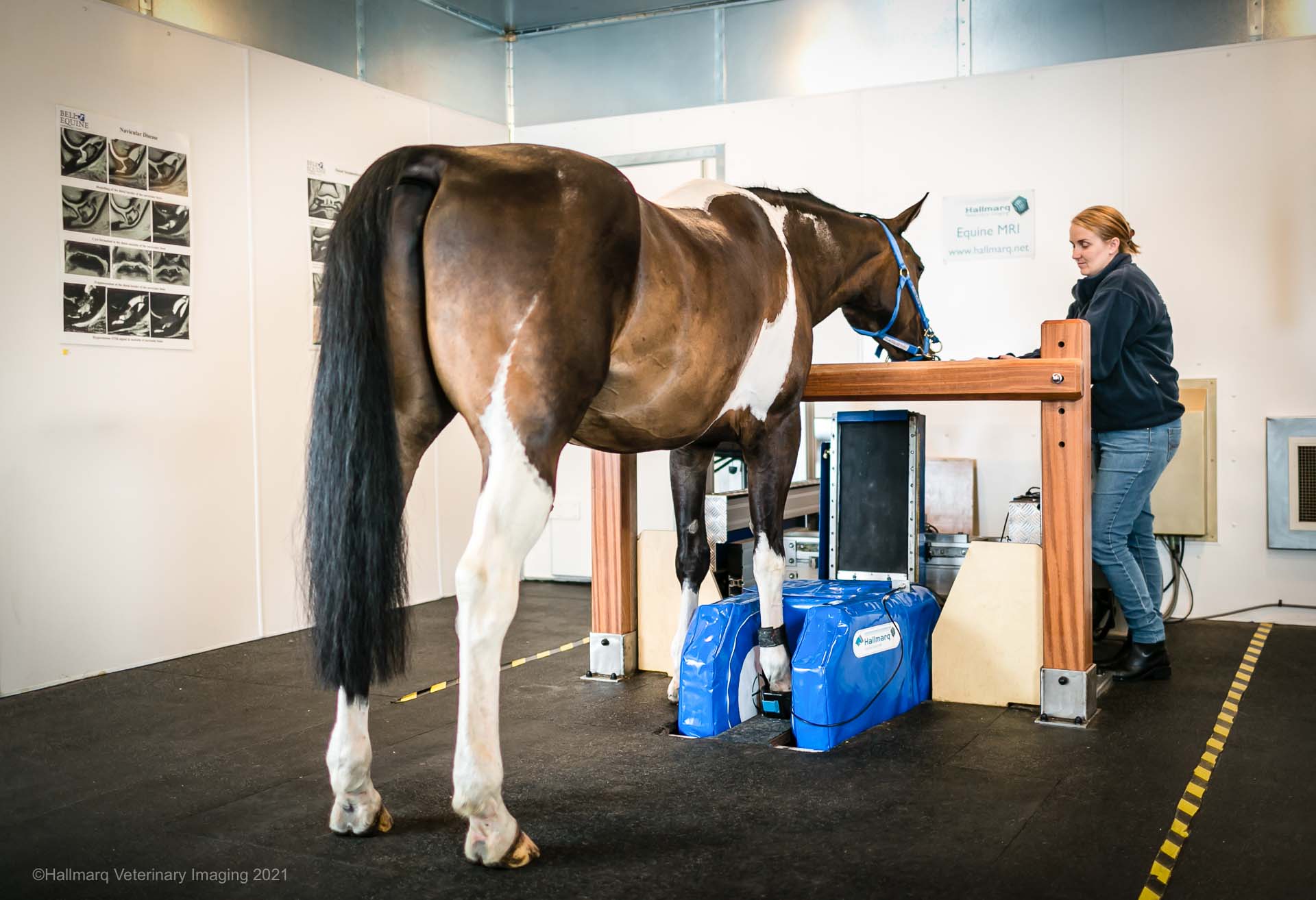
South Eastern Equine Hospital is excited to offer the gold standard in lameness diagnosis with the recent installation of the Hallmarq Standing MRI (sMRI). The module is specifically designed to help us deliver a safe, effective, and affordable diagnosis for your horse.
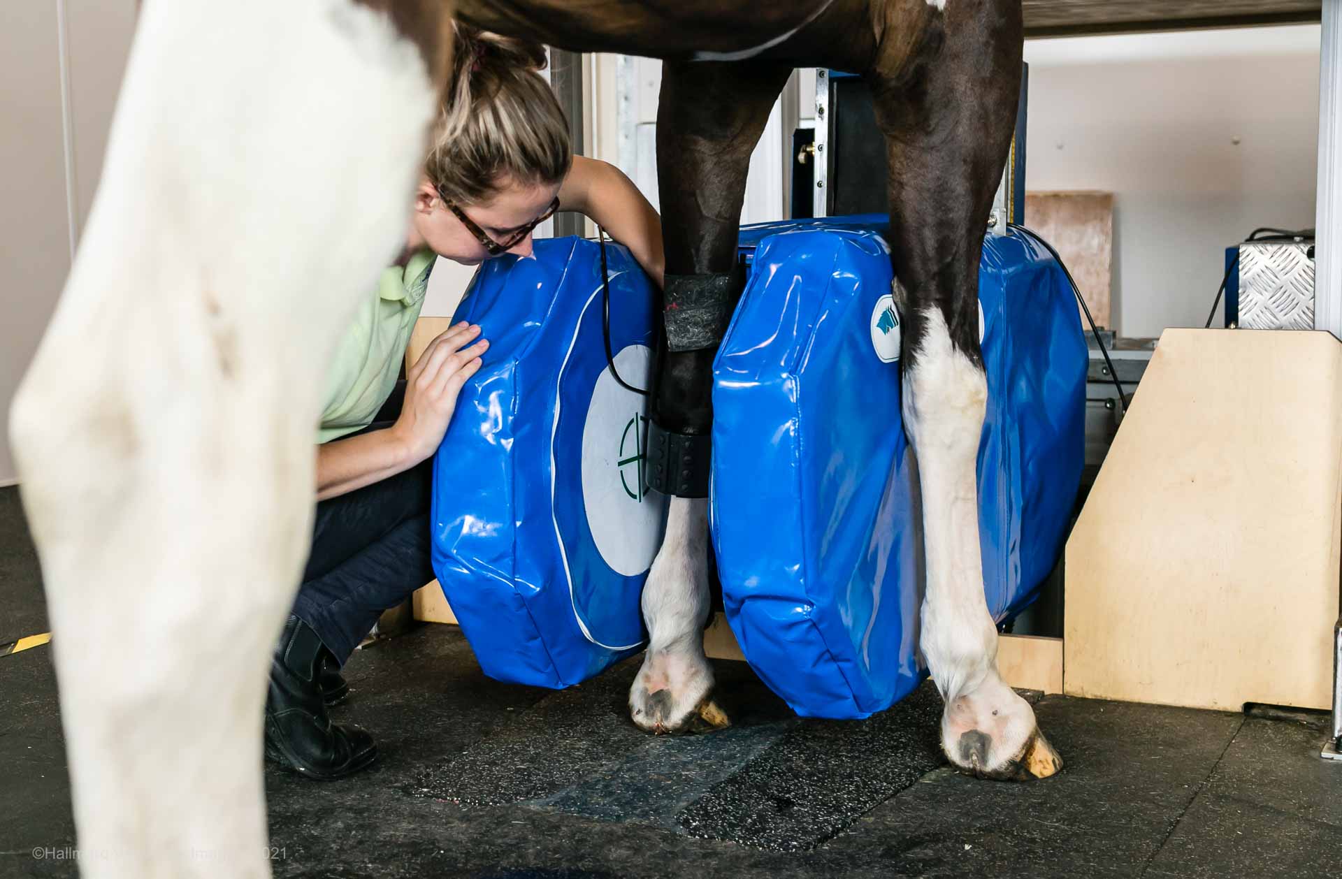
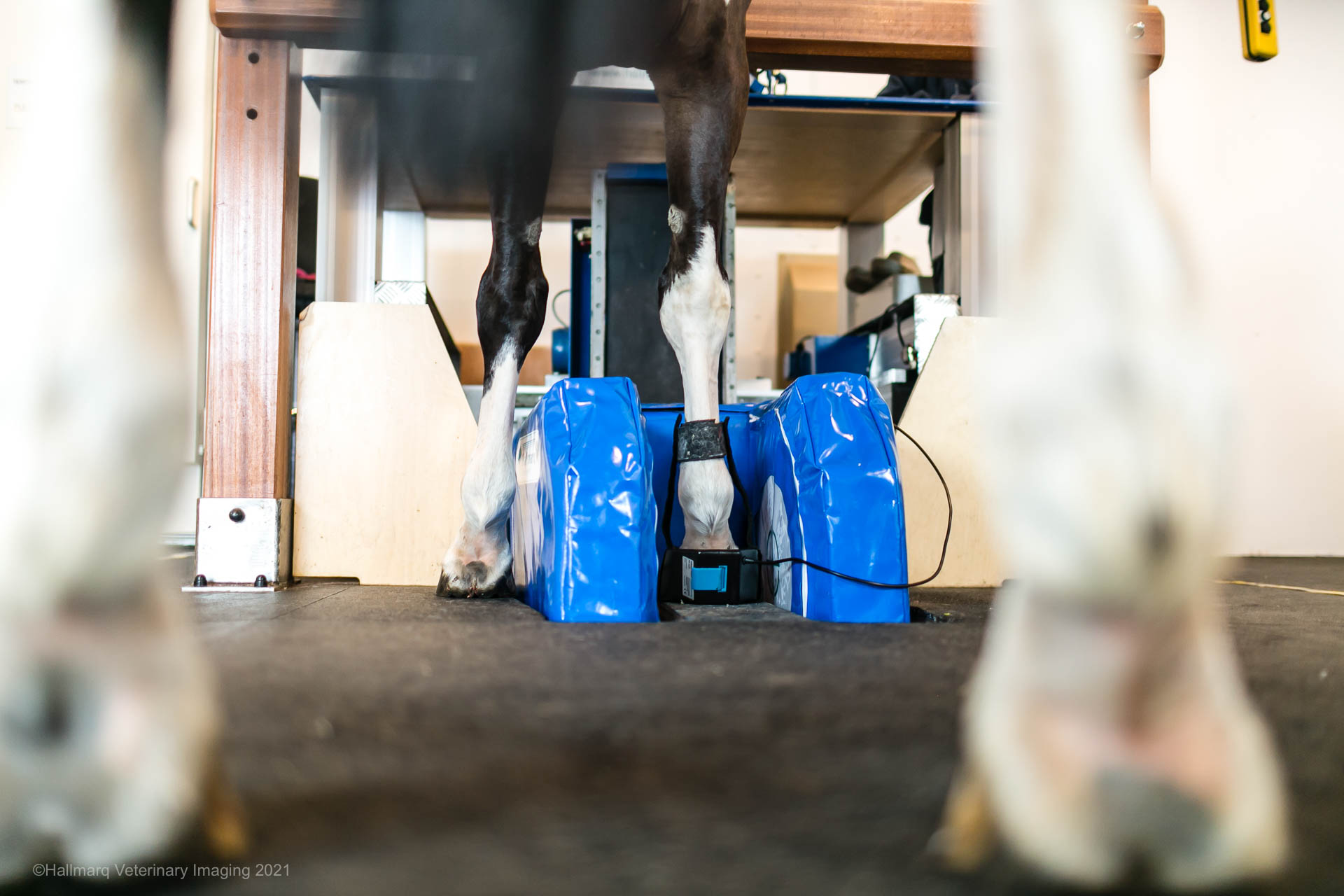
Standing MRI is a low-field procedure performed on the standing, sedated horse with foot, pastern, fetlock, carpus/tarsus, and even suspensory ligament studies available. MRI uses a magnetic field and a radio-frequency pulse enabling exceptional detail of soft tissue structures including tendons, ligaments, synovial structures, and cartilage. MRI scans also show lesions in bone and bone odema, that are not identified on radiographs.
At a Glance:
Length of stay: 2 nights
MRI type: Hallmarq Standing MRI
Areas imaged: Hoof to carpus and hock
General anaesthetic: No – performed under standing sedation
Preparation: Trot-up, shoe removal and foot radiographs the day before
Report turnaround: 48–72 hours
Image interpretation: Dr Alex Young, Specialist Veterinary Radiologist
Subsidy available: Racing Victoria MRI Subsidy (eligible horses only) https://www.racingvictoria.com.au/equine-welfare/veterinary-care/diagnostic-imaging-subsidy
Advanced imaging for an accurate diagnosis
South Eastern Equine Hospital is pleased to offer advanced standing MRI using the Hallmarq Standing Equine MRI system. Standing MRI is one of the most valuable diagnostic tools available for investigating persistent or complex lameness, allowing us to examine both bone and soft tissue structures in exceptional detail while your horse remains standing under sedation. It can image the lower limb from the hoof up to the carpus (knee) and hock.
Standing MRI avoids the risks associated with a general anaesthetic while providing detailed information that is often not visible on radiographs or ultrasound.
When is an MRI recommended?
Your veterinarian may recommend MRI when:
- Lameness has been localised to a specific region but the cause remains unclear.
- Radiographs (X-rays) or ultrasound have not provided a diagnosis.
- Soft tissue injuries are suspected within the hoof capsule or lower limb.
- A more accurate diagnosis is needed to guide treatment and prognosis.
- Previous treatments have not resolved your horse’s lameness.
An accurate diagnosis allows your veterinarian to develop the most appropriate treatment and rehabilitation plan for your horse.
What to expect
To ensure your horse is relaxed and well prepared, we ask that all MRI patients stay in hospital for two nights.
Day 1 – Admission and Preparation
Your horse will be admitted to South Eastern Equine Hospital and settled into one of our hospital stables.
On the day prior to the MRI, our veterinary team will perform:
- A comprehensive clinical examination.
- A trot-up and lameness assessment.
- Removal of shoes from the limb(s) to be scanned.
- Radiographs (X-rays) of the feet to ensure there are no retained metal fragments that could interfere with MRI image quality.
Completing these steps the day before the scan allows us to prepare your horse thoroughly and helps ensure the MRI appointment runs efficiently.
Day 2 – MRI Procedure
On the day of the MRI, your horse will be sedated before entering the MRI suite.
The scan is performed with your horse standing comfortably under sedation, avoiding the need for a general anaesthetic. Depending on the area being examined and the number of imaging sequences required, the MRI generally takes 1.5 to 3 hours.
Following the procedure, your horse will return to its stable where our veterinary and nursing team will continue to monitor its recovery overnight.
Day 3 – Discharge
Provided your horse is comfortable and no further procedures are required, they will be discharged to return home the morning after the MRI.
How are the images interpreted?
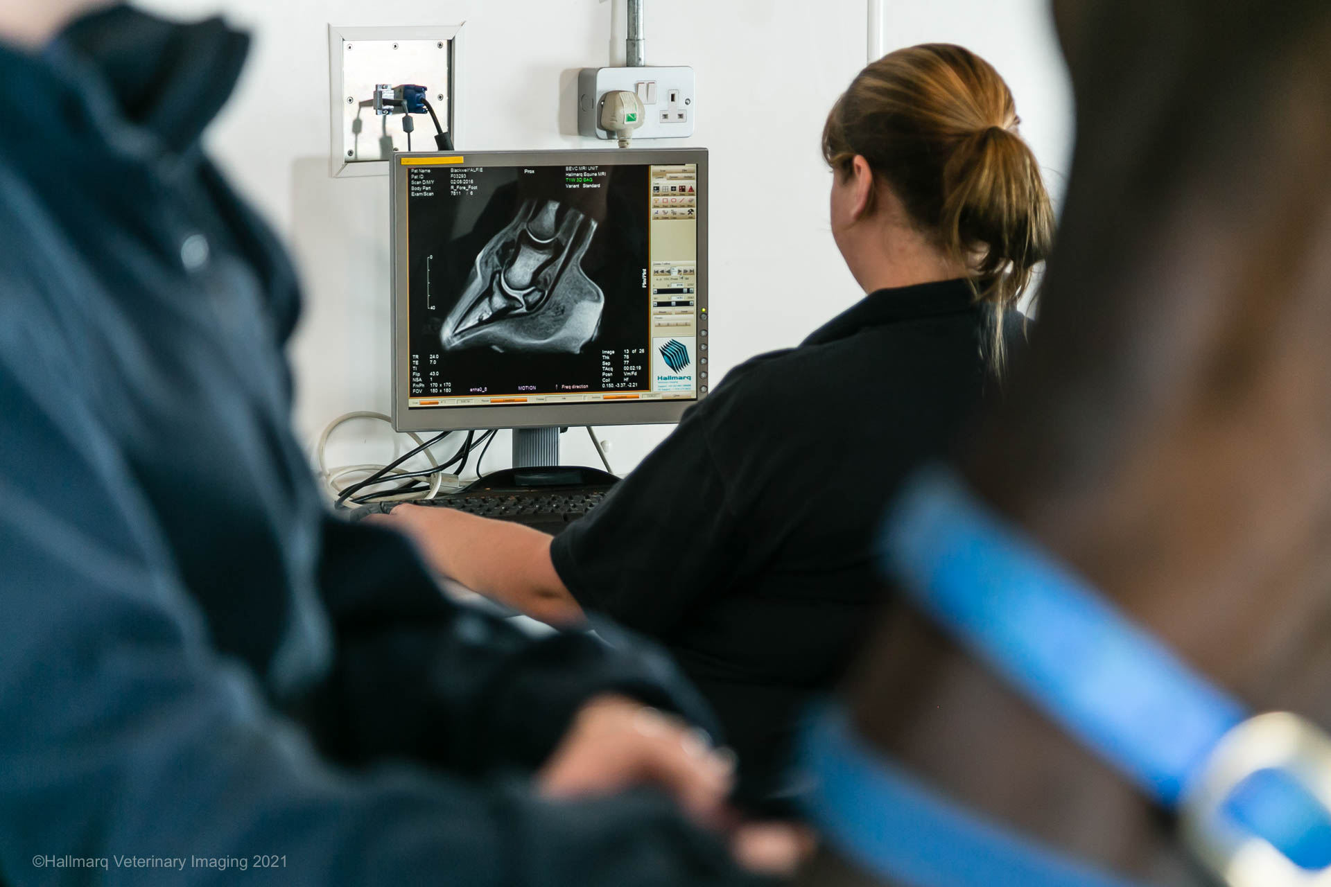
MRI produces hundreds of highly detailed images that require specialist interpretation.
All MRI studies are reviewed by Dr Alex Young BVSc (Hons I) DACVR, DACVR-EVI, Specialist Veterinary Imaging. Dr Young is a registered specialist in veterinary diagnostic imaging with extensive experience interpreting equine MRI studies.
A comprehensive written report is usually available within 48–72 hours of the scan.
Once the report has been completed, the findings will be discussed with your referring veterinarian, who will work with you to develop the most appropriate treatment and rehabilitation plan for your horse.
Working with your referring veterinarian
South Eastern Equine Hospital is proud to work in partnership with your regular veterinarian to provide advanced diagnostic imaging and specialist support.
MRI examinations are typically performed following referral from your veterinarian, who has an understanding of your horse’s history, previous investigations and current management. Before your appointment, we will liaise with your veterinarian to review any previous radiographs, ultrasound images and clinical findings to ensure the MRI examination is tailored to your horse’s individual needs.
Following the scan, all images are interpreted by Dr Alex Young BVSc (Hons I) DACVR, DACVR-EVI, Specialist Veterinary Imaging, and a comprehensive report is provided to your referring veterinarian within 48–72 hours.
Our veterinarians will discuss the MRI findings with your referring veterinarian, and together we will formulate recommendations for treatment, rehabilitation and any further investigations that may be required. Your referring veterinarian will remain your primary point of contact for ongoing care, ensuring continuity of treatment once your horse returns home.
We value the collaborative relationship we share with referring veterinarians and are committed to providing timely communication and the highest standard of specialist support for every patient.
Racing Victoria MRI Subsidy
South Eastern Equine Hospital is pleased to participate in the Racing Victoria MRI Subsidy Program.
Eligible Thoroughbred racehorses that meet the Racing Victoria criteria may qualify for a subsidy towards the cost of their MRI examination. This initiative aims to support the early diagnosis and management of orthopaedic injuries, helping to improve horse welfare and racing outcomes.
If you believe your horse may be eligible, please speak with our team when booking your appointment. We can advise on the current eligibility requirements and assist with the necessary documentation.
Frequently Asked Questions
Will my horse need a general anaesthetic?
No. Our Hallmarq MRI is a standing MRI, meaning your horse remains standing throughout the procedure under sedation. This significantly reduces the risks associated with general anaesthesia while allowing excellent quality diagnostic images.
Why do the shoes need to be removed?
Steel shoes and even very small metallic fragments within the hoof can create artefacts on MRI images, reducing image quality. Removing the shoes and taking screening radiographs beforehand ensures we obtain the clearest possible images.
Is MRI safe?
Yes. MRI uses a powerful magnetic field rather than ionising radiation. Your horse is closely monitored throughout the procedure by our experienced veterinary and nursing team.
How long before I receive the results?
A detailed specialist report is typically available within 48 to 72 hours. Your referring veterinarian will then discuss the findings, diagnosis and recommended treatment options with you.
Preparing for your appointment
Before your horse arrives, please:
- Bring any previous radiographs, ultrasound images or veterinary reports if these have not already been forwarded by your veterinarian.
- Inform us of any medications your horse is currently receiving.
- Follow any feeding or management instructions provided when your appointment is booked.
- Contact us if your horse develops illness or injury before the scheduled appointment.
Video Links
What is the process of a Standing Equine MRI?
How does Standing Equine MRI work?
Questions?
If you would like to know more about our MRI service or discuss whether your horse may benefit from advanced imaging, please contact South Eastern Equine Hospital. Our team is happy to answer any questions and guide you through the referral process.
Need for Information?
Link to Hallmarq Website:
https://hallmarq.net/products/equine-mri/


Opening Hours
Mon - Fri: 8:30am - 5:30pm
Sat: Emergencies only
Sun: Emergencies only
Emergencies: 24/7

Address
359 Belgrave-Hallam Road
Narre Warren North
VIC 3804

Book an Appointment
In the hospital or on-site, we make it easy to get expert care.

Staff
Meet our passionate team of equine health experts.

Services
See our full range of equine veterinary services.
Request a Booking
Our team will reply to confirm your appointment.
South Eastern Equine Hospital
Clinic Hours
Mon - Fri: 8:30am - 5:30pm
Sat-Sun: Emergencies only
Available 24/7 for Emergencies
Address
359 Belgrave-Hallam Road
Narre Warren North
VIC 3804